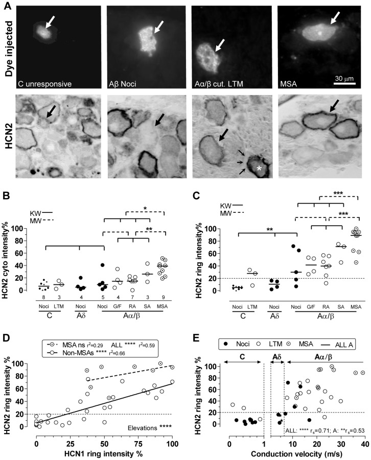
Figure 3. HCN2 immunointensity versus sensory properties and CV.
A:Top row dye-injected neurons; bottom row; the same neurons after ABC HCN2 immunocytochemistry; all with X40 objective. Large arrows indicate the dye-injected neurons. From left to right: C-unresponsive neuron shows cytoplasmic but no ring staining; the Aβ-nociceptor illustrated shows no, or a very weak, ring; Aα/β cutaneous LTM (field unit) with clear, not intense, ring; in the same field the asterisk shows a (non-dye-labelled) neuron identified as an MSA by strong staining on an adjacent section (not shown) for the α3 Na+ pump, which selectively labels MSAs. Also in that image, fine arrows show satellite cells unstained by HCN2. The right hand dye-injected neuron is a muscle spindle afferent (MSA), with a clear black ring. B: Cytoplasmic HCN2 relative intensities are elevated in MSAs versus other Aα/β neurons and all other neurons. C: HCN2 ring/edge staining is higher in MSAs than other Aα/β-neurons and than all other neurons; it is higher in Aβ-nociceptors than C-nociceptors, which showed no rings. Two out of three C-LTMs showed weak detectable ring staining. D: For non-MSA neurons ring intensities for HCN2 are highly significantly correlated with those for HCN1 (measured in different sections of the same neurons). For MSAs the linear regression was not significant (ns). The best linear regression line for MSAs was highly significantly elevated relative to non MSAs. E: HCN2 ring relative intensity is positively correlated with CV. For more detail and abbreviations see Fig. 2; for statistics see Methods.

