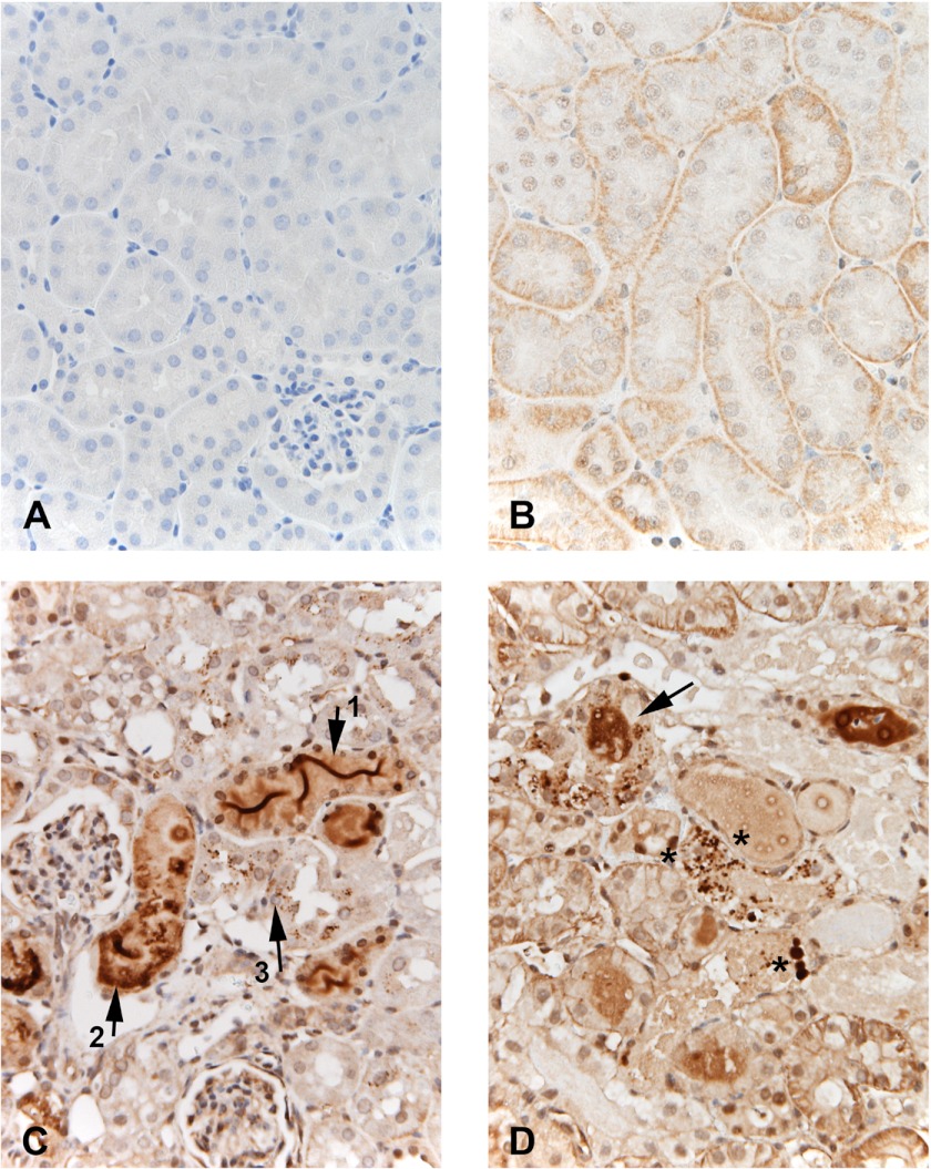
Fig. 6.
Immunohistochemical localization of Hpx following induction of CP and ischemic AKI. Compared with the isoytpe negative control (A), normal kidney (B) demonstrated trace Hpx staining with differing expression in different tubules. Staining appears predominantly at the basolateral membrane of proximal tubules. In contrast, the CP-treated kidney (C) demonstrated: 1) dense Hpx “ribbon like” staining within tubular lumina (arrow 1); 2) focal dense staining of tubule cytoplasm (arrow 2), and 3) numerous small Hpx containing reabsorption droplets within proximal tubules (arrow 3). Finally, the postischemic kidney (D) recapitulated the cisplatin findings with dense Hpx-stained casts (arrow) and prominent Hpx stained reabsorption droplets with proximal tubule cells (representative areas of increased uptake denoted by the asterisks). Lower asterisk denotes 4 prominent examples of this increased uptake.

