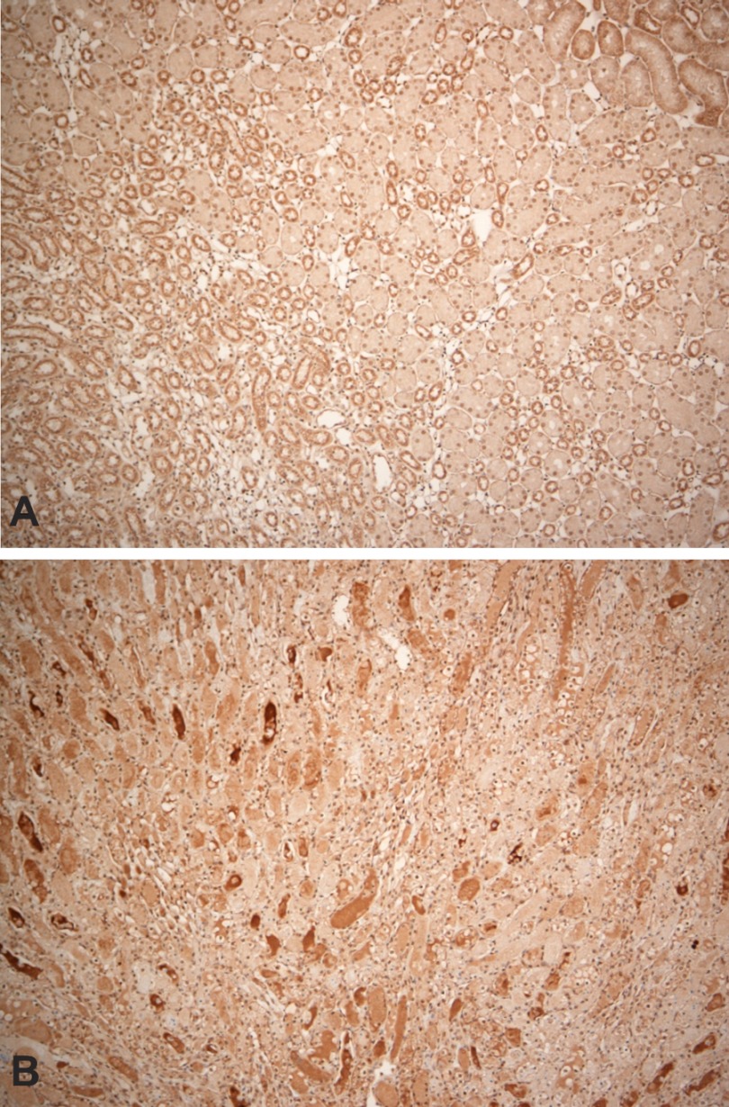
Fig. 7.
Immunohistochemical localization of Hpx within renal medulla in a control kidney (A) and in a kidney at 24 h postinduction of ischemic reperfusion injury. Compared with the control kidney (A), which shows minimal Hpx staining within the renal medulla, the postischemic kidney (B) manifests abundant Hpx staining casts. However, there was no increase in cytoplasmic Hpx staining and there was an absence of cellular Hpx-stained reabsorption droplets.

