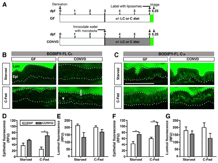
Figure 1. Fatty acids accumulate in the intestinal epithelium in the presence of microbiota and diet.

(A) Schematic of BODIPY-FL delivery assay in gnotobiotic zebrafish. Zebrafish derived germ-free (GF) at 0 days post-fertilization (dpf) were either reared GF (top) or inoculated at 3 dpf with normal microbiota (conventionalized, CONVD; bottom). From 3-6 dpf, fish were either starved, or fed a control (C) or low calorie (LC) diet (see Table S1). At 6 dpf, zebrafish were incubated with BODIPY-FL liposomes for 6 hrs and imaged or fixed for later imaging.
(B,C) Representative confocal images of the intestines of live 6 dpf GF and CONVD zebrafish incubated with BODIPY-FL C5 or C16. Scale bar, 50 μm.
(B) The intestinal lumen (Lum) and epithelium (Epi; bounded by dotted lines) of GF and CONVD zebrafish are indicated. The epithelium shows apical (white arrow) and basolateral accumulation of lipid droplets (white arrowhead) labeled with BODIPY-C5.
(C) Incubation with BODIPY-FL C16.
(D-G) Quantification of total epithelial (D,F) and luminal (E,G) fluorescence expressed in relative fluorescence units (RFU). Values represent the means ± SEM from 3 independent experiments: *, p<0.05; **, p<0.01.
