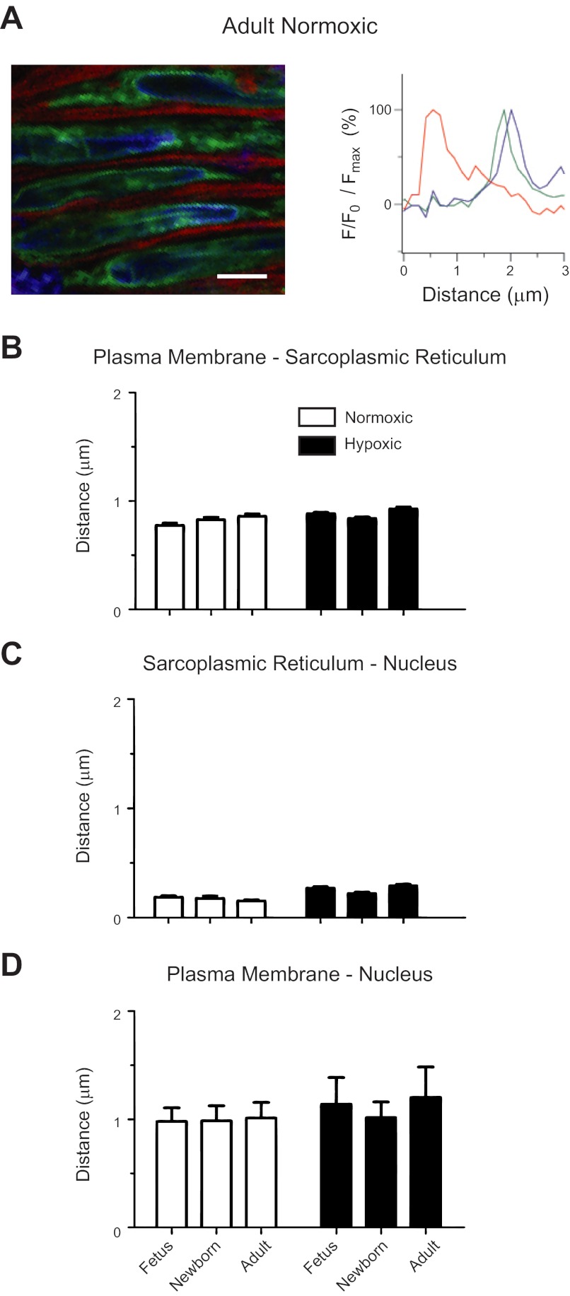
Fig. 1.
Ontogeny and long-term hypoxia (LTH) do not affect the spatial relationships between the sarcoplasmic-endoplasmic reticulum and the plasma membrane or nucleus. A: representative micrograph and associated intensity profiles of the plasma membrane (red), sarcoplasmic-endoplasmic reticulum (green), and nucleus (blue) of myocytes in a living pulmonary artery from normoxic adult, newborns, and fetuses. Bars indicate means ± SE for the distance between the sarcoplasmic endoplasmic reticulum and the plasma membrane (B) as well as nucleus (C–D). The difference in the distances for the various compartments in myocytes of fetal and adult pulmonary arteries of sheep shown in B, C, and D were within the calculated point-spread function for the Zeiss 710. Images were made in arterial segments from 4 fetal, 3 newborn, and 4 adult normoxic sheep and from 7 fetal, 4 newborn, and 6 adult long-term hypoxic animals with a ×63 water immersion c-Apochromat objective. F/F0: difference between peak spark fluorescence and background fluorescence. Scale bar = 5 μm.

