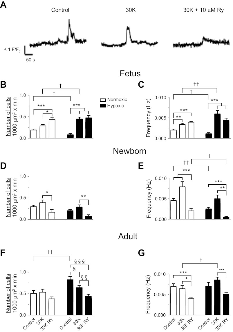
Fig. 8.
Effect of 30 mM K+ on whole-cell spatial and temporal Ca2+ signaling characteristics recorded in situ. A: baseline-subtracted fractional fluorescence (F/Fo) traces for Fluo-4 fluorescence are shown for individual pulmonary arterial myocytes from a normoxic adult sheep. Recordings were made in the absence (control) or presence of 30 mM K+ (30K) with or without 10 μM ryanodine (30K RY). Bars indicate means ± SE for normoxic (open) and long-term hypoxic (solid) conditions. B, D, and F: number of myocytes with Ca2+ responses each minute in a 1,000 μm2 area. C, E, and G: frequency of Ca2+ events *,§,†P < 0.05; **,§§,††P < 0.01; or ***,§§§P < 0.001, where the symbol * denotes significant difference within groups based on treatment using a Kruskal-Wallis 1-way ANOVA on ranks with a Dunn's multiple comparison procedure, the symbol § denotes significant difference within groups based on a 1-way ANOVA and an SNK multiple comparison procedure, and the symbol † denotes between groups based on their altitude using a Mann-Whitney U-test. Images were made with a ×60 Apochromat oil immersion objective.

