Fig. 9.
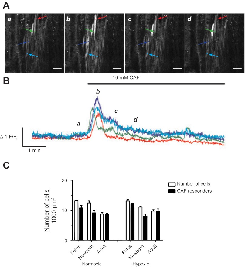
Caffeine causes whole cell Ca2+ responses in pulmonary arterial myocytes. A: representative fluorescent images at time points (a, b, c, and d) corresponding to the lower case italicized letters for the traces (B) of fractional Fluo-4 fluorescence shown for four pulmonary arterial myocyte from a LTH adult sheep. Colored arrows on images in A correspond to the placement of regions of interest for the traces of the same color in B. The 10 mM caffeine (CAF) was present during the time period denoted by the horizontal bar. C: number of myocytes in a 1,000 μm2 area measured in the multicolor images for Fig. 1 (open bars) and the number of myocytes with Ca2+ responses in the presence (solid bars) of 10 mM caffeine. Bars indicate means ± SE. No significant differences were noted between the number of myocytes responding to caffeine in each recording vs. the total number of myocytes in the arterial wall. Caffeine-elicited calcium responses were obtained in 3 fetal, 4 newborn, and 4 adult normoxic sheep and in 4 fetal, 4 newborn, and 3 adult LTH sheep. Images were made with a ×20 nonimmersion Apochromat or a ×63 water immersion c-Apochromat objective. Images in A were made with a ×63 objective. Scale bar = 20 μm.

