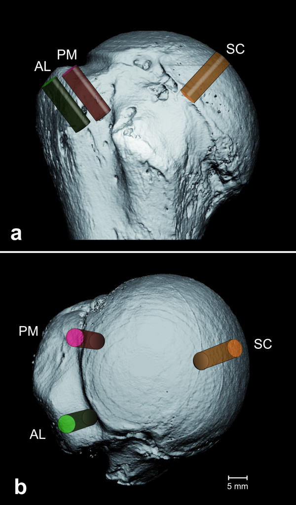
Figure 2.
Placement of the regions of interest.a) depicts an ap-view of the lesser tuberosity. Within the greater tuberosity, two rows were defined; one medial row adjacent to the articular surface, labeled with PM; and one lateral row along the lateral edge of the footprint, labeled with AL. Furthermore, a region was set into the subchondral region directly underneath the articular surface, marked with SC. b) gives an axial view of the humeral head with the lesser tuberosity on the bottom. The GT is divided into one posteromedial portion (PM) and one anterolateral part (AL).

