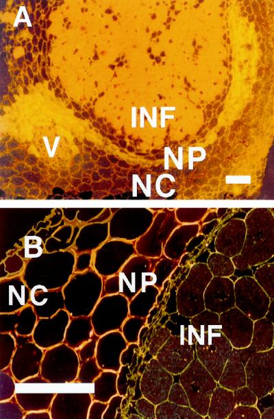
Figure 3.
Immunolocalization of AP in bean nodules (A) and negative control (B) using normal rabbit serum. Micrographs were taken using dark-field illumination, which makes Ag grains (AP) appear as bright dots. V, Vascular bundle. Other abbreviations are as in Figure 2. Each bar indicates 100 μm.

