
Figure 3. Intravascular release of doxorubicin.
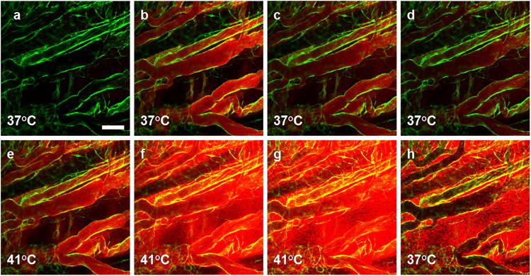
In the eNOS-GFP mouse model, GFP signal (green) is restricted to the endothelial cell layer. (a) Background vessels prior to injection. (b) Immediately after intravenous injection of Dox-TSL into tissue at 37°C, doxorubicin distribution becomes apparent by localized fluorescence (red) in the intravascular space (bolus phase, 0.5min). (c and d) 2-5min post-injection at 37°C, the drug fluorescence is still confined to the vascular space although the signal intensity is reduced, indicating a loss of liposomes from the blood stream. (e) 1min after the onset of heat treatment at 41°C, the intravascular drug signal increases due to intravascular release of doxorubicin and dequenching effects as the drug leaves the liposome interior. (f) After 5min of heating, extravascular doxorubicin accumulation becomes visible. (g) Drug accumulation increases in time with heating, and reaches a maximum at 15min into heating. (h) 10min after stopping heat most unbound free doxorubicin is washed out of the bloodstream and doxorubicin-stained nuclei remain fluorescent. Scale bar = 100μm.