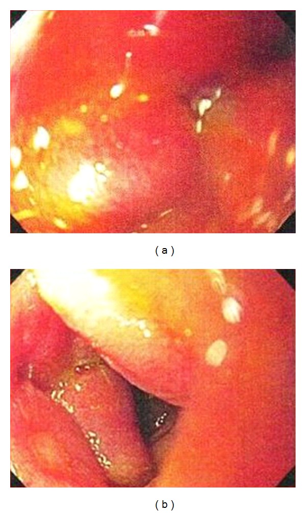
Figure 2.

Esophagogastroduodenoscopy image on the left shows edematous and eythematous mucosa of the 3rd portion of the duodenum. Image on the right shows an area of 5 mm of ulceration in the 4th portion of the duodenum.

Esophagogastroduodenoscopy image on the left shows edematous and eythematous mucosa of the 3rd portion of the duodenum. Image on the right shows an area of 5 mm of ulceration in the 4th portion of the duodenum.