Figure 5.
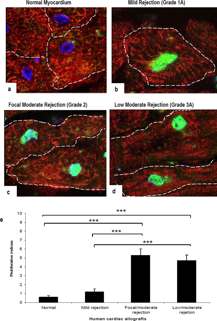
Confocal images of human cardiac biopsies. Nuclear staining for pH3 is illustrated in combination with α-sarcomeric-actin staining of the CM cytoplasm (green and red, respectively). (a). pH3 is absent in normal human myocardium. In comparison, a strong signal is apparent in CM in sections with mild grade 1A (b), focal moderate grade 2 (c), and low moderate grade 3A rejection (d). Proliferative indices (e); results are presented as means ± SEM. A significant difference is seen between normal myocardium and all three rejection episodes (***p<0.001) but not between grade 2 and 3A rejection. (Original Mags; ×63; Controls: n = 5; Allograft rejections, n= 5).

