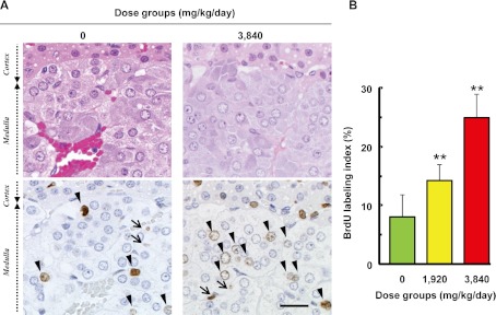
Fig. 1.
Stimulation of adrenal chromaffin cell proliferation in response to the serum calcium concentration in rats. A: HE staining (upper) and immunohistochemical staining of BrdU (lower) in the adrenal medulla of rats treated with or without GACa. BrdU-positive cells are found in the chromaffin cells (arrowhead) and endothelial cells (arrow), and BrdU-positive chromaffin cells are more frequently found in the GACa-treated groups than in the control group (0 mg/kg/day). Bar = 20 µm. B: The labeling index of the chromaffin cells in the adrenal medulla for each animal was calculated using the formula (BrdU-positive nuclei count) ÷ 500 (nuclei count) × 100. Values represent group means ± SD. Significant differences from the control are indicated with asterisks (** P<0.01). The BrdU labeling index was significantly and dose-dependently increased in the GACa-treated groups.

