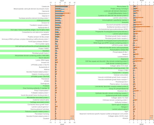
Fig. 3.
The secretome of H1 and H9 hESCs in MEF-conditioned medium was identified through MS analysis of the secretory pathway organelles. Only those proteins that were identified in both cell lines are listed. The NSAF values obtained in both H1 and H9 hESCs are plotted. Proteins that were not previously identified in hESCs are highlighted in green. Error bars denote standard deviation from triplicate measurements.

