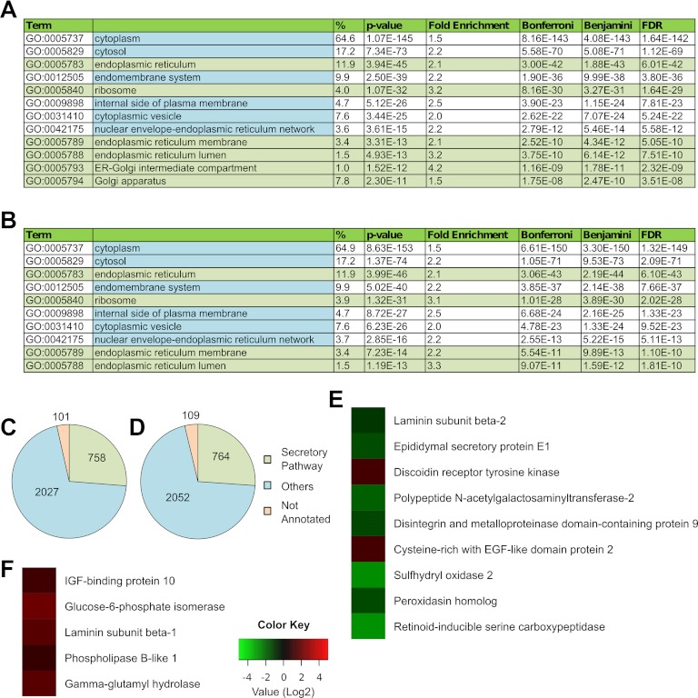
Fig. 4.
The proteome of hESCs changes in unconditioned medium. HESCs were exposed to unconditioned medium for 1 day, and secretory pathway organelles were isolated for MS analysis. GO enrichment analysis using DAVID shows statistically significant enrichment of the secretory pathway organelles from (A) H1 hESCs and (B) H9 hESCs. Distribution of proteins annotated to the secretory pathway organelles (endoplasmic reticulum, GO:0005783; endoplasmic reticulum-Golgi intermediate compartment, GO:0005793; ER to Golgi transport vesicle, GO:0030134; Golgi apparatus, GO:0005794; secretory granule, GO:0030141; transport vesicle, GO:0030133; and ribosome, GO:0005840) shown for (C) H1 hESCs and (D) H9 hESCs. The protein NSAF values obtained from these cells were compared with those obtained from H1 and H9 hESCs grown in MEF-CM. Statistically significant differences in NSAF values of secretory proteins are shown for (E) H9 hESCs and (F) H1 hESCs.

