Fig. 1.
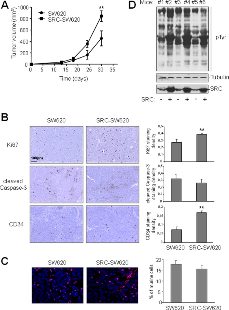
SRC increases tumor growth and pTyr content in CRC xenograft models. A, SRC promotes growth of xenograft tumors in nude mice. The mean tumor volume (in mm3) ± S.E. (n = 5) obtained after injection of parental SW620 cells (control, circle) or SW620 cells that over-express SRC (SRC-SW620, square) is presented over time. B, SRC promotes cell proliferation and angiogenesis in xenograft tumors. Representative sections (left-hand panels) and quantification (right-hand panels) of immunohistochemical analysis (left) showing cell proliferation (anti-ki67), apoptosis (anti-cleaved Caspase 3), and angiogenesis (anti-CD34) in xenograft tumors derived from SW620 and SRC-SW620 cells as described under “Experimental Procedures.” C, SRC expression does not affect stromal cell infiltration in tumors. A representative example (left) and quantification (right) of the percentage of mouse cells included in xenograft tumors derived from SW620 and SRC-SW620 cells. Tumor sections were labeled via in situ hybridization with a mouse-specific probe as described under “Experimental Procedures.” The merge between mouse labeled nuclei (red) and DAPI-stained nuclei (blue) is shown. D, SRC increased pTyr content in tumors. Western blotting of total tumor lysates from mice injected with SW620 (−) or SRC-SW620 (+) cells using the anti-phosphotyrosine 4G10 antibody. The levels of Tubulin and SRC are also shown. *p < 0.05 and **p < 0.01 using Student's t test.

