Fig. 1.
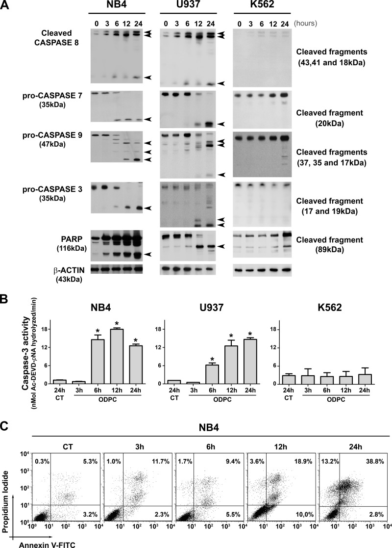
Kinetics of apoptosis induced in leukemia cells by treatment with ODPC. A, apoptosis was initiated within 6 h of exposure to 25 μm ODPC in AML cell lines. Antibodies were used to detect activated caspases, pro-caspase, or cleaved fragments. B, colorimetric caspase-3 activity assay using the specific peptide substrate (AcDEVD-pNA) for activated caspase-3. Measurements were carried out using 30 μg total protein; p < 0.05 compared with control (analysis of variance). C, annexin V and propidium iodide staining was measured via flow cytometry of NB4 cells. The K562 cell line showed a pattern of resistance compared with the U937 and NB4 cell lines (A and B). On the basis of these results, we used NB4 cells treated with 25 μm ODPC for 3 h to perform the proteomic screening. Arrows indicate the cleaved fragments.

