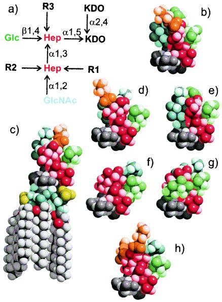
FIG. 1.
(a) Structural representation of seven inner core galE LPS structures of the N. meningitidis immunotypes. (b to h) Three-dimensional space-filling molecular models of the galE mutant inner core structures color coded as follows: PEtn (orange), Glc (green), GlcNAc (pale blue), heptose (Hep) (red), 2-keto-3-deoxyoctulosonic acid (KDO) (dark grey), and lipid A (pale grey). Molecular models were constructed using a Metropolis Monte Carlo approach and are drawn with the same orientation for the KDO residue. The lipid A moiety is shown in panel c. (b) L2 galE (R1 = PEtn-6, R2 = Glc-α1,3, R3 = H); (c) L3 galE (R1 = H, R2 = PEtn-3, R3 = H); (d) L4 galE (R1 = PEtn-6, R2 = H, R3 = H); (e) L5 galE (R2 = Glc-α1,3, R1 = H, R3 = H); (f) MC58 lpt3 galE (R1, R2, R3 = H); (g) 1000 galE (R1 = H, R2 = H, R3 = Glc-β1,2); and (h) 2220Y galE (R1 = PEtn-6, R2 = PEtn-3, R3 = H). L2-16 was raised to structures b and d, LPT3-1 was raised to structure f, and L3B5 was raised to structure c (see Materials and Methods).

