Fig. 3.
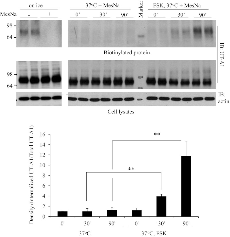
UT-A1 internalization assay. UT-A1 MDCK cells were first biotinylated and then rewarmed at 37°C in the presence or absence of FSK for the different times. The noninternalized biotin on the cell surface was stripped by sodium 2-mercaptoethane sulfonate (MesNa) treatment. The cells were lysed in RIPA buffer and the total lysates were used for Western blot with UT-A1 and actin antibodies. Internalized proteins were recovered by streptavidin beads and processed for Western blotting with UT-A1 antibody. The bands were quantified (n = 3). The internalized UT-A1 was normalized to the UT-A1 from total lysates. The relative density of the control (time point 0) was set as 1. **P < 0.01.

