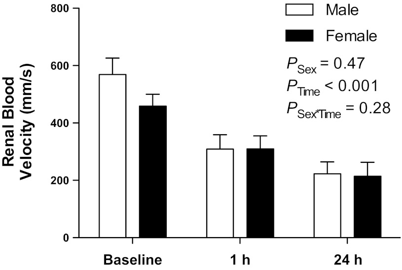
Fig. 3.
Peak systolic renal blood velocities measured by pulsed-wave Doppler ultrasound in the right kidney of male (n = 7) and female (n = 6) mice before 1 h ischemia (baseline) and following 1 and 24 h reperfusion. Data are presented as means ± SE and were analyzed by two-factor ANOVA, testing for main effects of sex (PSex), time (PTime), and the interaction between the two (PSex×Time).

