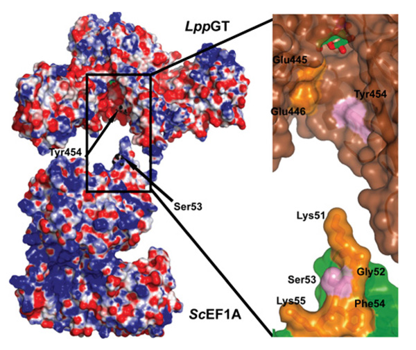
Figure 3. Electrostatic surface representation of LppGT and ScEF1A.
Left-hand panel: LppGT has a negatively charged binding groove, which may interact with the positively charged loop on ScEF1A (PDB code 2B7B [37]) that carries the acceptor serine. Tyr454, in the putative binding groove on LppGT, and the acceptor serine (Ser53) on ScEF1A are indicated by arrows. Right-hand panel: higher magnification representation of the putative interaction site between LppGT and ScEF1A. The surface of LppGT and ScEF1A are represented in brown and green respectively. Tyr454 and Ser53 are shown as sticks in pink; charged residues are in yellow (including some other residues forming the loop in which Ser53 is localized, such as Gly52 and Phe54).

