Figure 1.

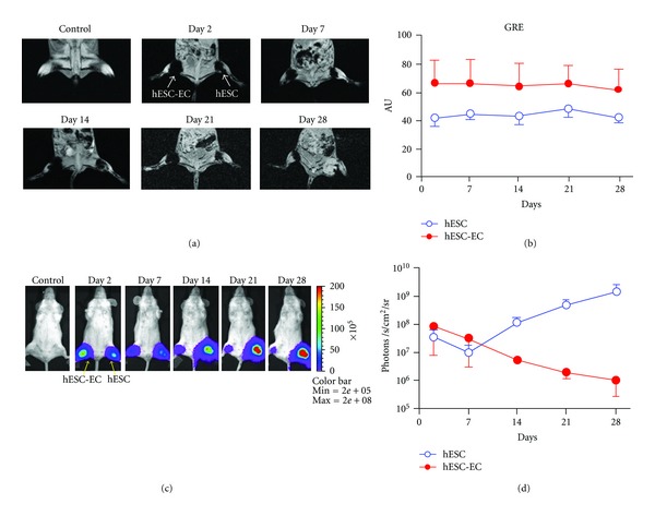
Direct comparison of reporter gene imaging (genetic labeling) versus iron particle imaging (physical labeling) for tracking stem cells. (a) Human ESCs were cultured under normal conditions or on gelatin/fibronection-coated plates to induce endothelial cell differentiation. These predifferentiated human ESC-derived endothelial cells (hESC-ECs) and undifferentiated ESCs were SPIO-labeled (with Feridex) and 1 × 10⁶ cells were then injected into mouse hindlimbs. MR images of one representative animal show the cells at days 2, 7, 14, 21, and 28. (b) MR does not show survival differences between the two groups, as the signal is steady throughout all imaging timepoints, with a higher signal in the hESC-EC group through day 28. (c) These same ESCs were transduced with the human ubiquitin promoter driving fireflyluciferase(Fluc) and enhanced green fluorescence protein (eGFP). These cells were then cultured as in (a) prior to transplantation into the hindlimb of a mouse. (d) BLI showed divergent survival profiles for the two groups, with proliferation of ESC and acute donor cell death of pre-differentiated hESC-ECs. This study demonstrated that MRI provided detailed information on the anatomical location of cells, but not on cell viability. Reporter gene imaging is a better indicator of cell viability and proliferation [36].
