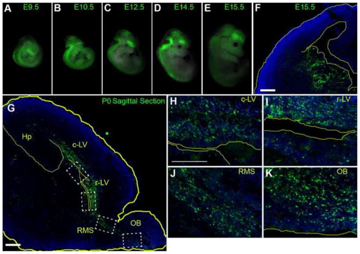
Figure 4. CR2-GFP+ cells are predominant in the embryonic ganglionic eminence and in VZ/SVZ of the mouse neonatal cortex.
(A–E) Whole mount images of transgenic mouse embryos at various stages: E9.5 (n=3), E10.5 (n = 3), E11.5 (n = 4), E12.5 (n = 4), E14.5 (n = 5), and E15.5 (n=10). GFP expression was exclusive to the CNS ranging from the telencephalon to the sacral spinal cord. (F) Fluorescent microphotographs of coronal brain sections of the transgenic mouse at E15.5 showing CR2-GFP+ cells were predominantly located in the VZ/SVZ of the ganglionic eminences (GE). (G–K) CR2-GFP+ cells in saggittal brain sections of transgenic mouse at P0 were primarily located in regions surrounding the lateral ventricle, the olfactory bulb, the rostral migratory stream, and hippocampus. Blue represent nuclear marker Dapi staining. c-LV = caudal lateral ventricle, Hp = hippocampus, r-LV rostral-lateral ventricle, SVZ = subventricular zone, VZ = ventricular zone, OB = olfactory bulb, rms = rostral migratory stream. Scale bars = 20 μm.

