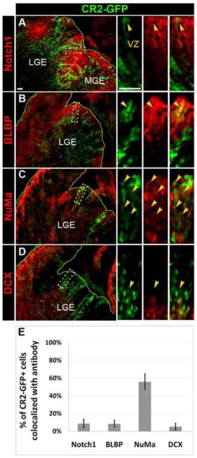
Figure 5. CR2-GFP+ cells are in neural progenitors undergoing asymmetric division in E15.5 transgenic mouse.

(A–D) Fluorescent microphotographs of coronal brain sections of the transgenic mouse at E15.5 showing CR2-GFP+ cells co-stained with radial glial markers, Notch1 (A) and BLBP (B), a marker for asymmetric division, NuMa (C), and an early neuronal marker, DCX (D). (E) Histograph showing the percentage of CR2-GFP+ cells co-localized with cell-specific markers. Less 10% of CR2-GFP+ cells were co-stained with Notch1 (9±5%), BLBP (8±4%), and DCX (5±4%). 56±9% of CR2-GFP+ cells were co-stained with NuMa. Scale bar = 20 μm.
