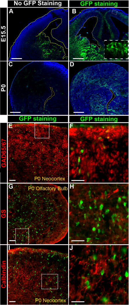
Figure 7. Immunostaining recovered CR2-GFP+ cells are GABAergic interneuron progenitors in the transgenic neocortex.
Fluorescent microphotographs of coronal brain sections of the transgenic mouse at E15.5 (A–B) and P0 (C–D). (A) At E15.5, without anti-GFP staining, the CR2-GFP+ cells were prominently seen in the VZ/SVZ of the GE. (B) With anti-GFP staining, three additional populations of CR2-GFP+ cells were revealed: the ventral ganglionic eminence (GE) below the VZ/SVZ, the basal (upper) layers of the neocortex, and VZ/SVZ of the neocortex. (C) At P0, without anti-GFP staining, CR2-GFP+ cells were only seen mostly VZ/SVZ surrounds the lateral ventricle of the ventral forebrain, and a few GFP+ cells in the cortical plate. (D) With anti-GFP staining, more than twice as many CR2-GFP+ cells were revealed, and they were located in the VZ/SVZ of the entire lateral ventricle and widely spread over the cortical plate. (E-J) Sagittal brain sections of P0 transgenic mouse were co-stained with anti-GFP antibody and cell specific markers, GAD65/67 (E–F), glutamine synthetase (GS) (G–H), and Calbindin (I–J). The majority of immunostained GFP+ cells were co-stained with GAD65/67 but not with GS or calbindin. Scale bars = 20 μm.

