
Figure 1.
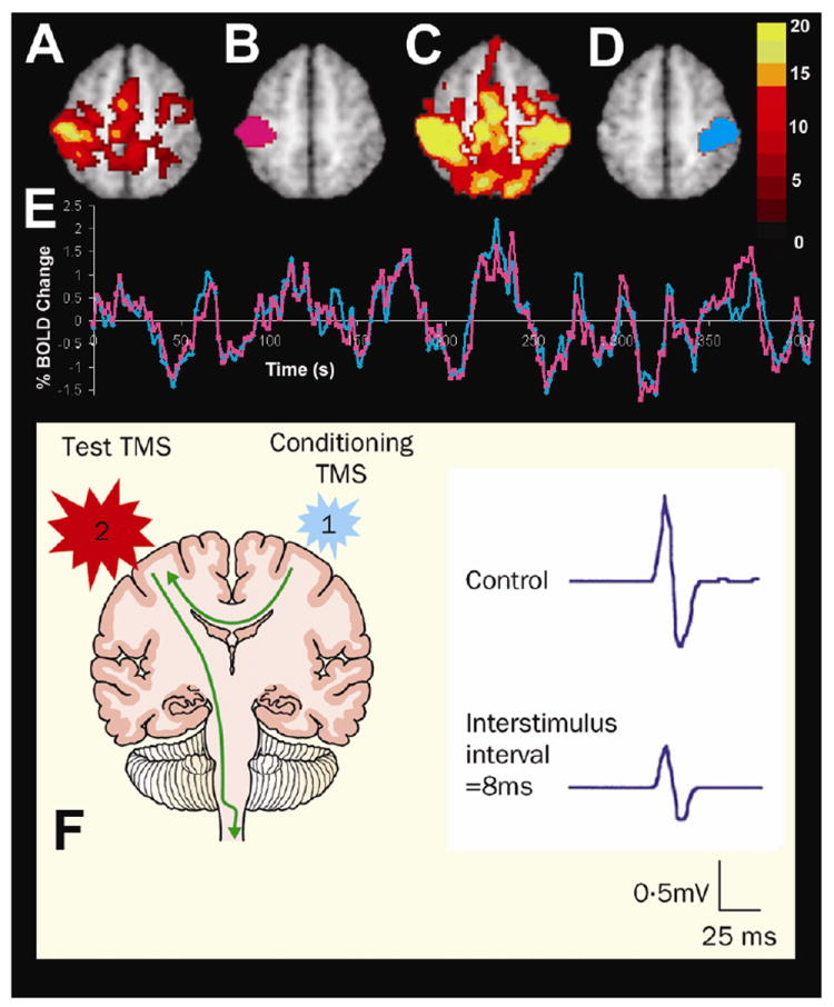
Connectivity between the motor cortices assessed with resting state functional connectivity MRI and dual-coil stimulation with TMS. The top panel shows fMRI activation in response to a right hand button press (A), a left somatomotor region of interest (B), resting state functional connectivity with this left somatomotor cortex region of interest (C), a right somatomotor cortex region of interest defined on the basis of the resting state functional connectivity (D), and spontaneous fluctuations recorded in the left (pink line) and right (blue line) somatomotor cortices during the resting state conditions showing significant interhemispheric correlation (modified with permission from (Fox, Snyder et al. 2007)). The lower panel shows the effect of transcallosal inhibition using dual-coil TMS. When a conditioning pulse is delivered to the left motor cortex 8 ms before the test pulse is delivered to the right motor cortex the motor evoked potential recorded from the left hand is significantly decreased (modified with permission from (Kobayashi and Pascual-Leone 2003)).