
Figure 1.
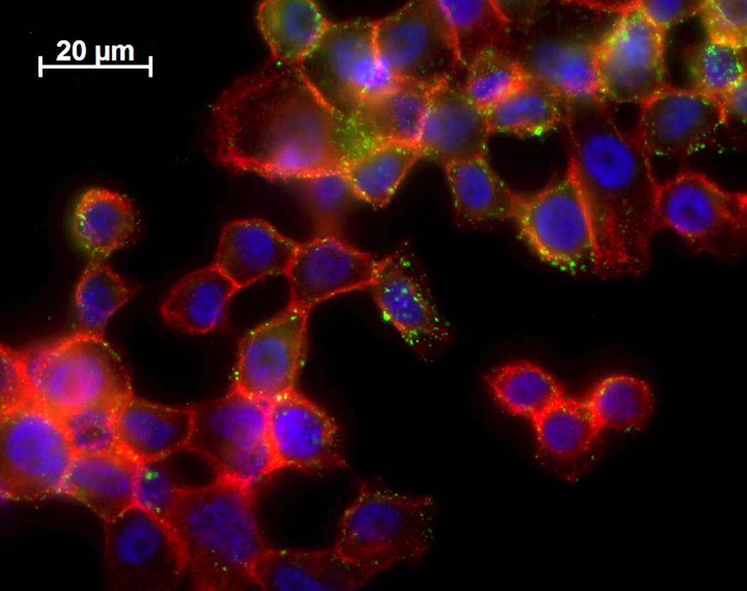
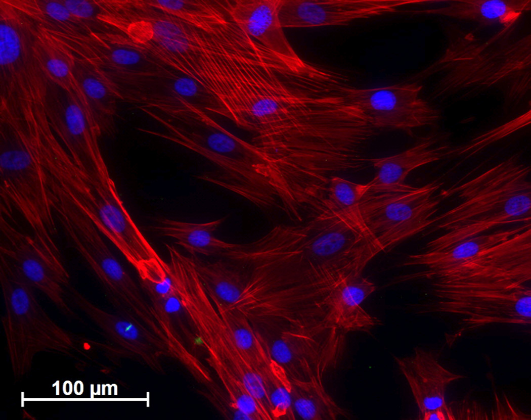
Microscopy of FR-α for A) HeLa and B) fibroblasts. Folate receptor was detected with primary antibody using anti-FR-α mouse IgG and secondary antibody of anti-mouse IgG conjugated to FITC (green). In addition, Hoechst stained was used to detect the nuclei (blue) and rhodamine phalloidin stained the cells’s cytoskeleton (red).