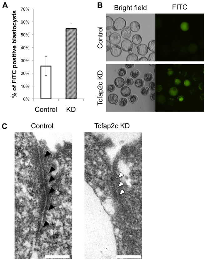
Fig. 4.

Disruption of TJ complexes and paracellular sealing in Tcfap2c KD embryos. (A) A 4 kDa FITC-conjugated dextran was used to test the permeability of TJs in Tcfap2c KD and control blastocysts. A difference in permeability was observed between Tcfap2c KD and control blastocysts. Error bars indicate mean ± s.e.m. (B) Representative brightfield and fluorescence images of Tcfap2c KD and control blastocysts subjected to the FITC-dextran assay. (C) TEM analysis of TJ complexes in Tcfap2c KD and control morulae. At 90 hph, several electron-dense plaques (black arrowheads) were observed at apical cell-to-cell contact sites in control embryos. By contrast, in Tcfap2c KD embryos the electron-dense regions (white arrowheads) at the apical cell-to-cell contact site were absent. Scale bars: 200 nm.
