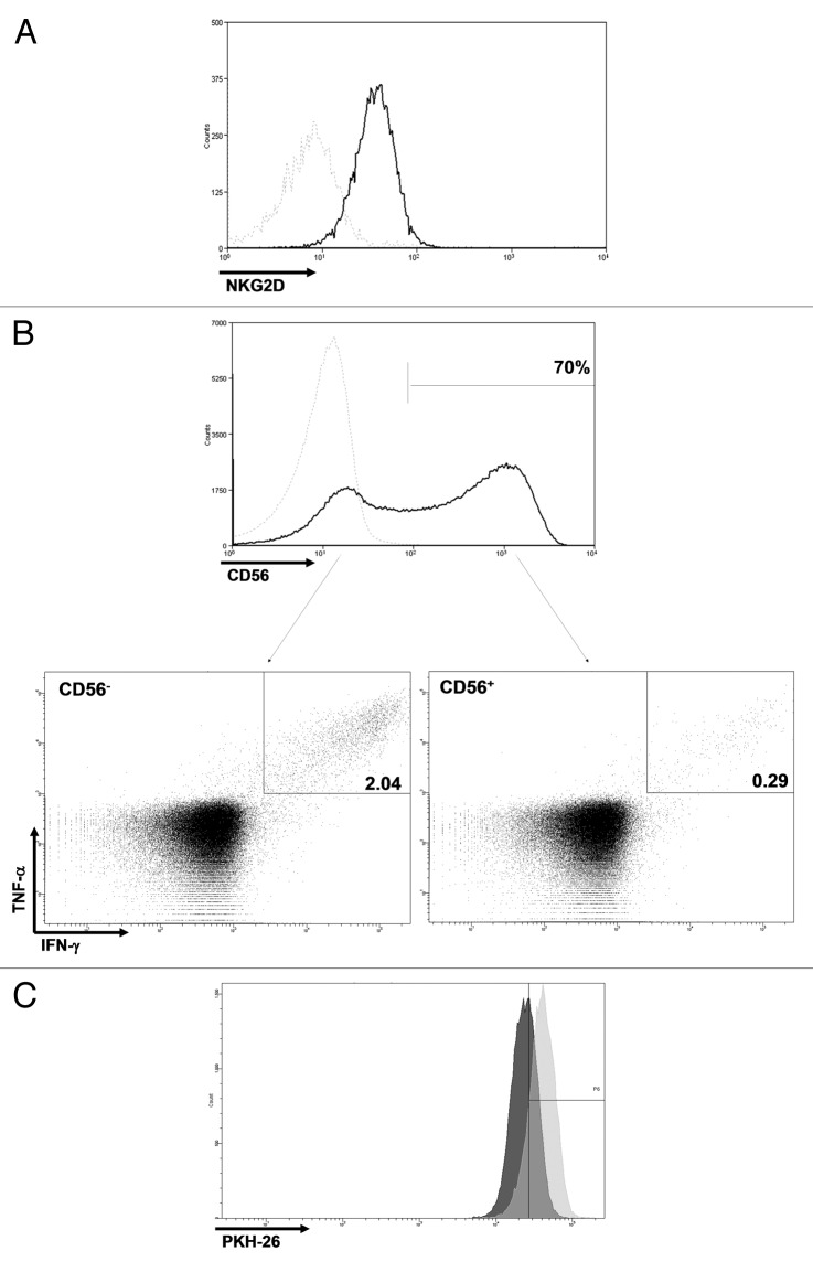
Figure 3. Phenotypic and proliferative characteristics. (A) Vδ1 T cells expressed NKG2D. (B) CD56 was expressed by a large fraction of Vδ1 T cells, and cells with in vitro anticancer activity were enriched in the CD56- compartment. Dotted light gray line: isotype control. (C) From day 8 to day 10 of the rapid expansion protocol (REP), undivided cells were 85% of αβ T cells vs. 45% of Vδ1 T cells. Light gray, αβ T cells; dark gray, Vδ1 T cells.

An official website of the United States government
Here's how you know
Official websites use .gov
A
.gov website belongs to an official
government organization in the United States.
Secure .gov websites use HTTPS
A lock (
) or https:// means you've safely
connected to the .gov website. Share sensitive
information only on official, secure websites.
