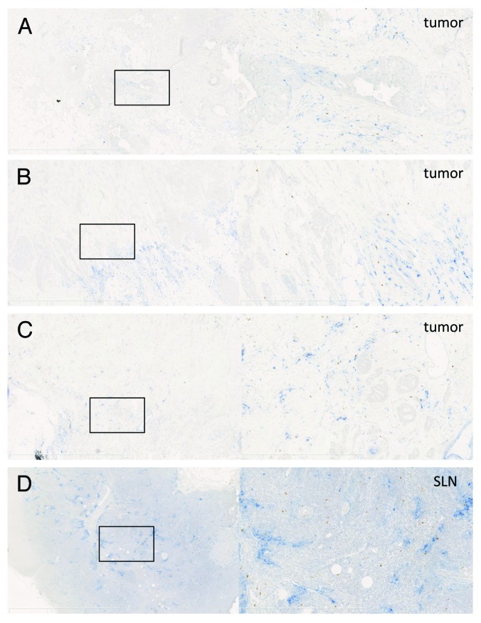
Figure 5. Arginase 1 staining in tumors and tumor-draining lymph nodes. (A–D) Examples of CD14 (blue, nuclear hematoxilin is light blue) and ARG1 (brown) staining in breast tumors (A–C) and a tumor-draining lymph node (D). Left panels = 2.5× magnification, right panels = 10× magnification of the selection on the left side.

An official website of the United States government
Here's how you know
Official websites use .gov
A
.gov website belongs to an official
government organization in the United States.
Secure .gov websites use HTTPS
A lock (
) or https:// means you've safely
connected to the .gov website. Share sensitive
information only on official, secure websites.
