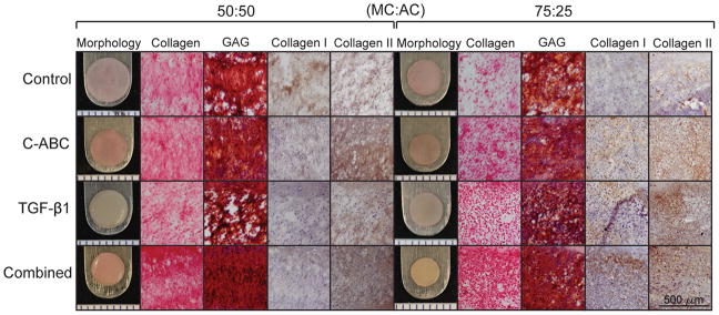
Fig. 1.
Gross morphology, histology and IHC of constructs at t = 5 wk. Fibrocartilage was treated with either C-ABC alone, TGF-β1 alone, the two agents combined, or left untreated (control). Collagen was stained for using picrosirious red, GAG was stained for using Safranin O/Fast Green, while IHC was used to stain for collagen types I and II. Collagen and GAG staining was stronger in the 50:50 constructs, with the combined treatment producing constructs with the most uniform staining in both cell ratios. IHC staining showed different patterns of collagen types I and II between cell ratios as well as among bioactive treatments within each cell ratio. Scale bar for histology and IHC images is 500 μm and the markings on the morphology images are 1 mm apart.

