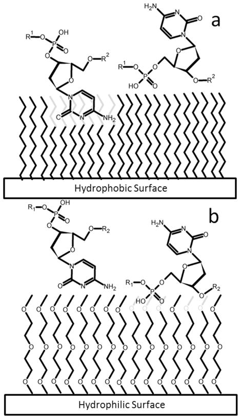
Figure 1.
A schematic diagram of hypothetical surface interactions for the nucleobase cytosine (left molecule) and the phosphate backbone (right molecule). The surfaces represent (a) a non-hydrogen-bonding hydrophobic surface, and (b) a non-hydrogen-bonding hydrophilic surface, All chains in the surface layer are the same length; the “shorter” apparent chains are intended to demonstrate immersion/intercalation of the adsorbate within the monolayer.

