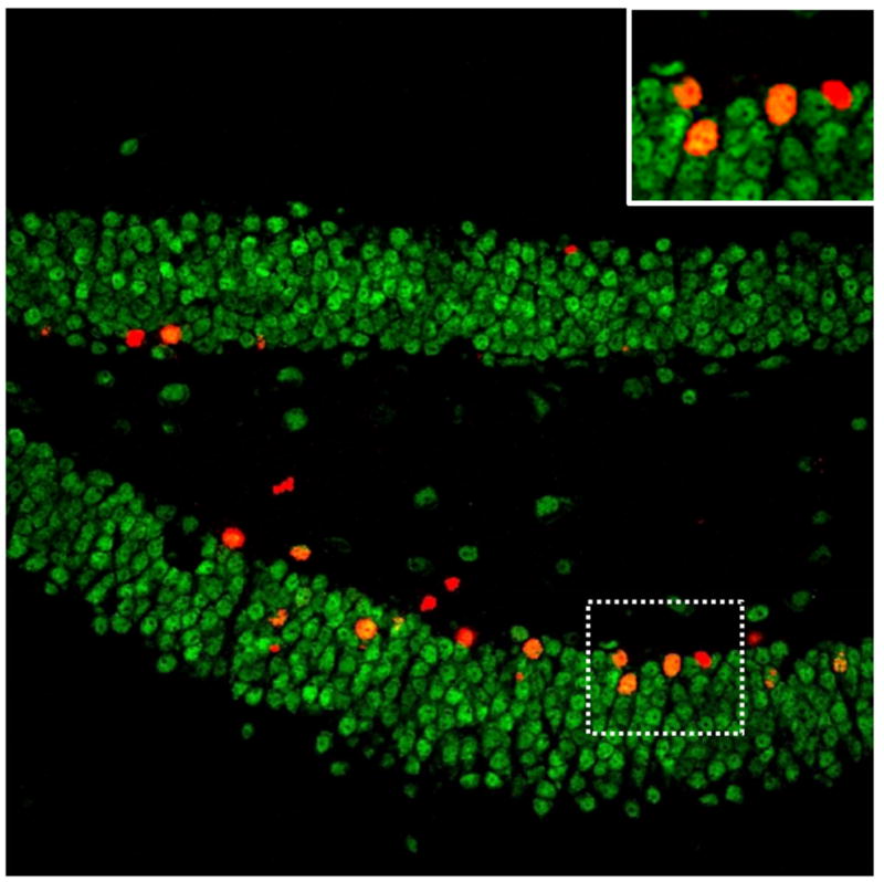
Figure 1.

Image of a mouse dentate gyrus double-labeled with fluorescent antibodies against BrdU (new cells in red) and NeuN (mature neuronal marker in green). New cells that have differentiated into neurons are co-labeled with both BrdU and NeuN (new neurons in orange). The area within the dashed lines contains 3 new neurons and one new cell of unknown phenotype and is shown zoomed in at the top right.
