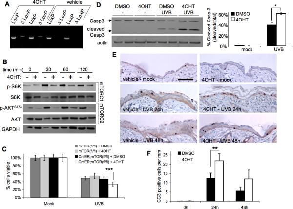
Figure 5. Induction of mTOR deletion in mouse primary keratinocytes and epidermis sensitizes cells to UVB-induced apoptosis.
K5-CreERT2;mTORfl/fl and mTORfl/fl keratinocytes were harvested from 1-3 day-old pups and cultured in 4OHT (1mM) or vehicle for 3 days prior to UVB (50mJ/cm2) exposure. A, PCR analysis of K5-CreERT2;mTORfl/fl primary keratinocyte DNA harvested 24 h after final 4OHT treatment. ΔLoxP denotes primers specific to the recombined mTOR allele. B, Immunoblot analysis of mTORC1 and mTORC2 activation markers in K5-CreERT2;mTORfl/fl keratinocyte extracts harvested following UVB (50mJ/cm2) exposure. C, MTS cell viability (mean ± SEM) of mTORfl/fl and K5-CreERT2;mTORfl/fl keratinocytes at 24h post UVB; *** p < 0.005. D, Immunoblot analysis (K5-CreERT2;mTORfl/fl primary keratinocytes) of caspase-3 and quantification (mean ± SEM) at 9-h post UVB; * p<0.05. A-D data are representative from 2-4 independent experiments. E, Representative cleaved Caspase-3 staining images of skin sections (scale bar = 200μm) from K5-CreERT2;mTORfl/fl mice treated topically with 4OHT and exposed to UVB (120mJ/cm2). F, Quantification of cleaved capsase-3 (CC3) staining (mean ± SEM) for 3-5 mice/group; ** p < 0.01.

