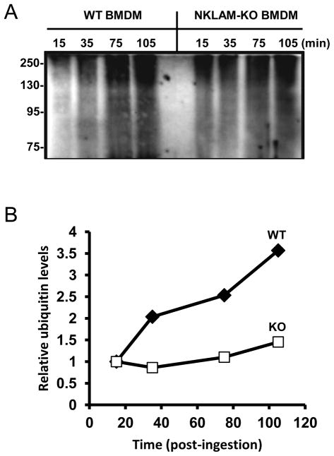
Figure 4. Ubiquitination of phagosome proteins during maturation.
(A) WT and NKLAM-KO BMDM were stimulated with LPS/IFNγ for 18 hr. The cells were then incubated with IgG-opsonized magnetic beads at 4°C for 10 min. After incubation at 37°C for the times indicated, the phagosomes were isolated and the phagosome proteins were immunoblotted for ubiquitin. (B) Densitometry was performed on each lane. Graph represents the ratio of each lane relative to the 15 min time point for WT (closed diamonds) and NKLAM-KO (open squares). The immunoblot and graph are representative of five independent experiments.

