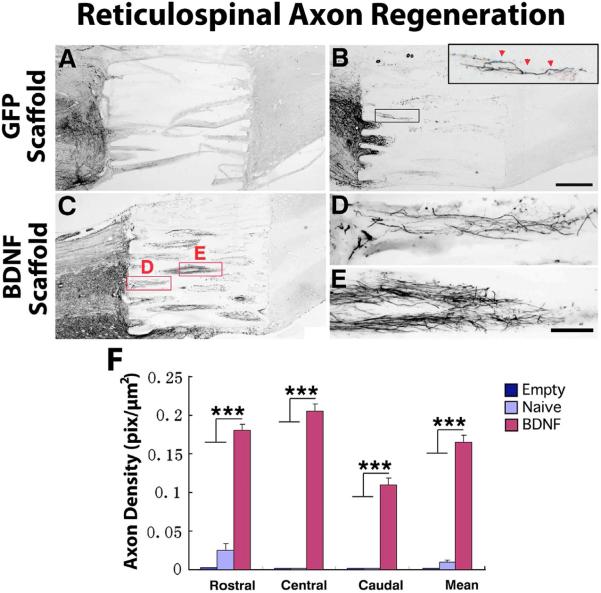
Figure 2. Regeneration of BDA-labeled Reticulospinal Motor Axons into Templated Agarose Scaffolds in Complete Transection Sites.
(A) Empty scaffolds contained very few regenerating reticulospinal axons and these extended for only short distances in the lesion site. In all panels rostral is to the left. Such scaffolds, nearly devoid of regenerating axons, tended to distort on histological processing. (B) Scaffolds filled with a cellular matrix of bone marrow stromal cells expressing the reporter gene GFP, but not expressing a growth factor, exhibited modest axonal penetration of scaffolds but these failed to extend to distal aspects of the lesion site (inset). (C) Scaffolds filled with BDNF-secreting marrow stromal cells exhibited significant increases in the total number of axons entering scaffolds and in total axons reaching the distal aspect of the lesion site, quantified in panel F. Higher magnification views of (D) axonal entry into scaffold channels, and (E) midway through lesion, are shown at higher magnification. Axons invariably extended in linear trajectories. However, axons did not regenerate into host spinal cord tissue beyond the lesion site. (F) Quantification of axonal density in scaffolds at short (0–500μm), middle (750–1250μm) and caudal (1500–2000μm) aspects of channels. Kruskall Wallis test, χ2 p<0.01; Dunn post-hoc with Bonferroni correction ***p< 0.001. Asterisks indicate comparisons of BDNF group to both Empty and Naïve groups. Error bars indicate SEM. Scale bar: A–C 500μm; D, E 100μm.

