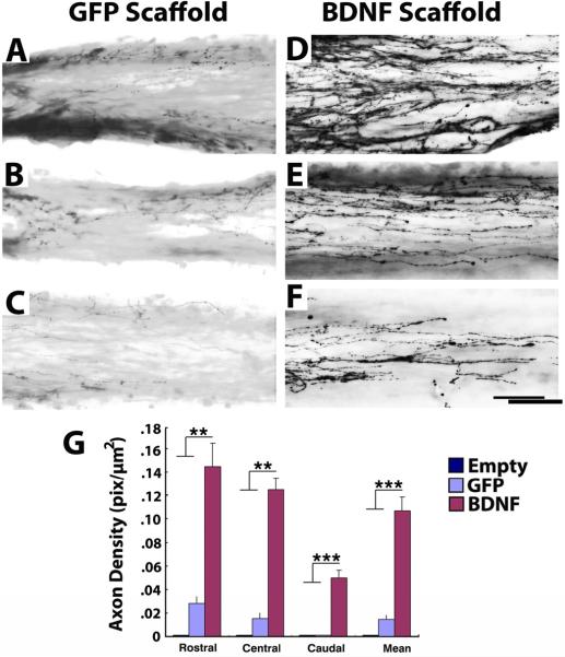
Figure 3. Regeneration of Serotonergic Axons into Templated Agarose Scaffolds in Complete Transection Sites.
(A–C) Modest numbers of serotonergic axons labeled for 5HT penetrate scaffolds filled with GFP-expressing marrow stromal cells. A, entry into scaffold; B, mid-point of scaffold; C, caudal aspect of channel. (D) Significantly greater numbers of serotonergic axons penetrate scaffolds filled with BDNF-secreting marrow stromal cells, quantified in G. D, axonal entry into channel; B, mid-point of scaffold; C, caudal aspect of channel. Axons extended in linear trajectories. (G) Kruskall Wallis test, χ2 p<0.01; Dunn post-hoc with Bonferroni correction **p<0.01, ***p< 0.001. Asterisks indicate comparisons of BDNF group to both Empty and Naïve groups. Scale bar: A–C, 500μm; E–I, 50μm.

