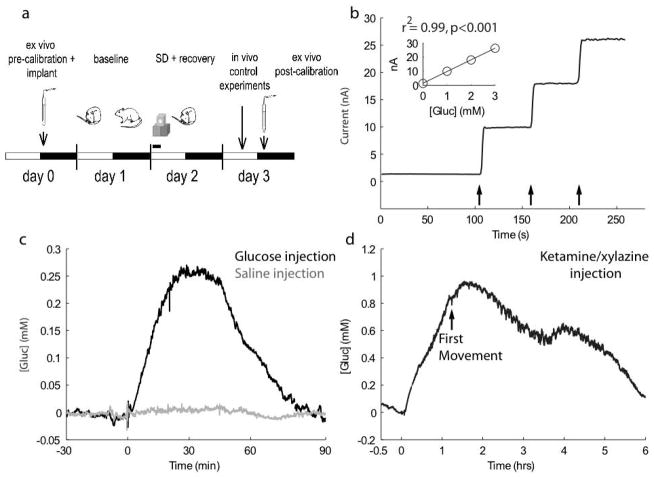
Figure 1. In vitro and in vivo assessment of glucose-sensitive microelectrodes.
a) Schematic of experimental design. White and black bars indicate the light and dark phase, respectively. b) A typical in vitro calibration conducted following removal of electrode from a chronic in vivo recording. Arrows depict 1mM additions of glucose to the calibration solution. After 3 days of in vivo recordings, this electrode still responds robustly (8.27 nA/mM) and linearly to glucose, as particularly evident in the inset. c) [Gluc] in frontal cortex largely increases following an i.p. injection of glucose (black; 2mL of 30% glucose) but is largely unaffected by an equivalent volume injection of saline (grey). d) [Gluc] in frontal cortex increases during ketamine/xylazine anesthesia (87 mg/kg and 13 mg/kg). Following recovery from anesthesia, [gluc] remains elevated for ~5 hours.

