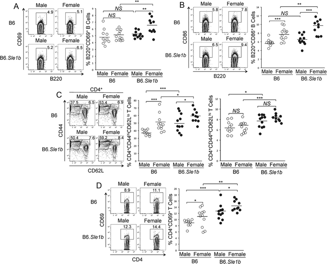
Figure 1. Increased spontaneous activation of B cells and CD4 helper T cells in B6.Sle1b female mice.
Flow cytometric analysis was performed on splenocytes from 6–9 month old, sex-matched B6 and B6.Sle1b mice after staining for activated B cells using mAbs against B220, CD69 and CD86. The percentage of B220+CD69+ (A) and B220+CD86+ (B) activated B cells are shown in rectangular gates (left panels) and in scatter plots (right panels). Activated CD4 T cells were analyzed using antibodies against CD4, CD44, CD62L and CD69. The percentage of CD4+CD44hiCD62Llo short-lived effector and CD4+CD44hiCD62Lhi effector memory helper T cells are shown in rectangular gates (left panel, C) and in scatter plots (middle and right panels, respectively, C). The percentage of CD4+CD69+ activated T cells are shown in rectangular gates (left panel, D) and in scatter plot (D, right panel). Each circle represents an individual mouse and horizontal bars represent the mean values. Statistical analysis was performed as described in Materials and Methods.

