
Figure 6. Significantly higher number of proliferating B cells in B6.Sle1b female mice.
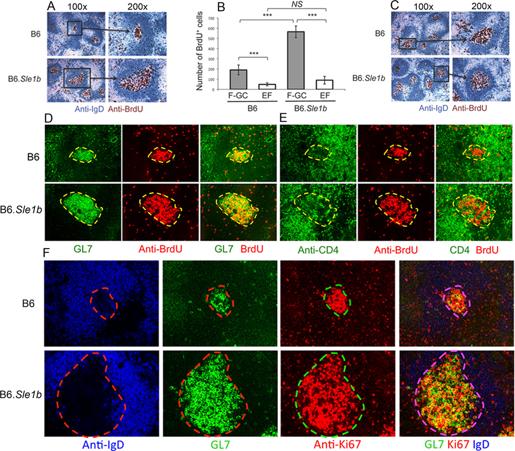
(A) Spleen sections from 6–9 month old B6 and B6.Sle1b mice injected with BrdU 12 hours and 1–2 hours prior to sacrifice were stained for anti-IgD (blue) and anti-BrdU (brown). Original magnification of the images was 100× (left) and 200× (right). (B) Semi-quantitative analysis of the number of BrdU+ cells in follicular-GC (F-GC, gray bars) versus extrafollicular (EF, white bars) regions. BrdU+ cells were counted in two randomly picked areas per spleen at ×100 original magnification. Five B6 and 5 B6.Sle1b female mice were analyzed. (C) Spleen sections obtained from SRBC-immunized 5–6 week old female B6 and B6.Sle1b mice were analyzed for BrdU+ cells inside the GCs as described in A. Parallel spleen sections were obtained from 6–9 month old female B6 and B6.Sle1b mice and stained for either GL7 (green) and anti-BrdU (red) as shown in (D) or anti-CD4 (green) and anti-BrdU (red) as shown in (E). (F) Spleen sections from 6–9 month old female B6 and B6.Sle1b mice were stained for anti-IgD (blue), GL7 (green), and anti-Ki67 (red). Original magnification of the images in C-E was 200×. These data were generated from at least 5 female mice of each genotype.