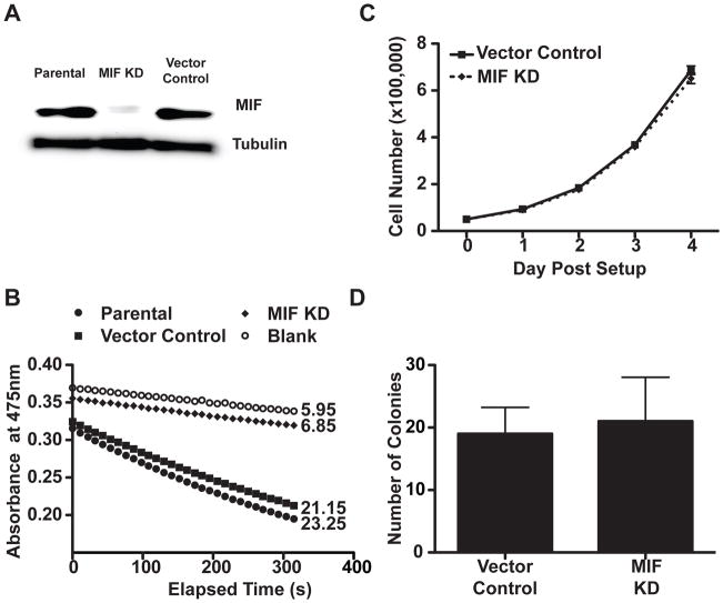
Figure 1. MIF KD cells exhibit similar grow properties in vitro.
A, Immunoblot demonstrates effective MIF depletion in the MIF KD cells compared with the vector control and the parental 4T1 cells. Lysates from cell lines were normalized by Bradford assay for total protein concentration, run on a gel, and the membrane was probed using anti-MIF and anti- tubulin antibodies. B, Effective MIF depletion is confirmed by loss of tautomerase activity. Cell lysates normalized for total protein were assayed for tautomerase activity using a colorimetric substrate. The reaction was monitored for decolorization over 5 minutes at 475nm. Velocities (rate of decolorization/unit time) are indicated to the right of each kinetic plot. C, Depletion of MIF does not alter in vitro proliferation. 5×10⁴ cells per well were plated in 6-well dishes. At the indicated time points, cells were trypsinized and counted. D, Depletion of MIF does not alter anchorage independent growth of 4T1s. 4×10⁴ cells were seeded in soft-agar in a 6 well plates. The plates were incubated at 37°C for 2 weeks, then the colonies counted in three randomly selected fields. For panels C and D, n=3 and each is representative of 3 independent experiments.

