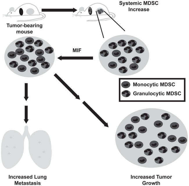
Figure 8. Proposed model of the role of MIF in cancer progression.
Tumor bearing animals demonstrate a systemic increase in myeloid derived suppressor cells, detectable both in the blood and the spleen. The majority of these cells are of the granulocytic phenotype. Within the tumor, high levels of MIF promote the abundance of the more suppressive monocytic phenotype, correlating with an increase in tumor growth and metastasis in the presence of an intact immune system.

