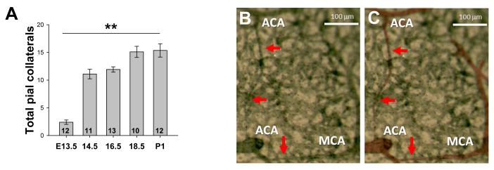
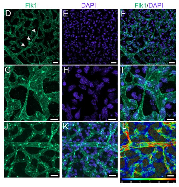
Figure 1. Most pial collaterals form between E13.5 and E14.5 from sprout-like extensions from established arterioles.
A, Time-course of collateral formation (total, all collaterals between ACA and MCA of both hemispheres of CD1 mice; **p<0.01; number of embryos from > 2 litters at base of bars). B, Collaterals form superficial to the pre-existing pial capillary plexus, are not yet tortuous, and taper as they extend from the parent vessel. C, Arterioles in B pseudocolored for clarity. D-F, Branches eminate from distal-most arterioles (arrowheads), course superficial to the plexus, and appear to be led by a tip cell. G-I, High magnification image of above tip cell. J-L. Depth coding on an image stack shows that collaterals form superficial to the plexus. Scalebars: A-C, J-L:20μm, D-F:10μm.


