
Fig. 3. Characterization of Ly6Chi and Ly6Clo macrophages accumulating in the liver following APAP intoxication.
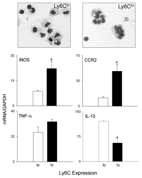
Liver nonparenchymal cells, isolated 48 h after treatment of wild type mice with APAP, were stained with anti-CD11b and anti-Ly6C antibodies, as described in the Materials and Methods. CD11b+ cells were sorted based on expression of Ly6C into CD11b+/Ly6Chi and CD11b+/Ly6Clo subpopulations. Upper panel. Cytospin preparations of sorted cells were stained with Giemsa. Lower panel. mRNA expression of sorted cells was assessed by RT-PCR. Data were normalized relative to GAPDH; bars represent mean ± SE (n=4-6).