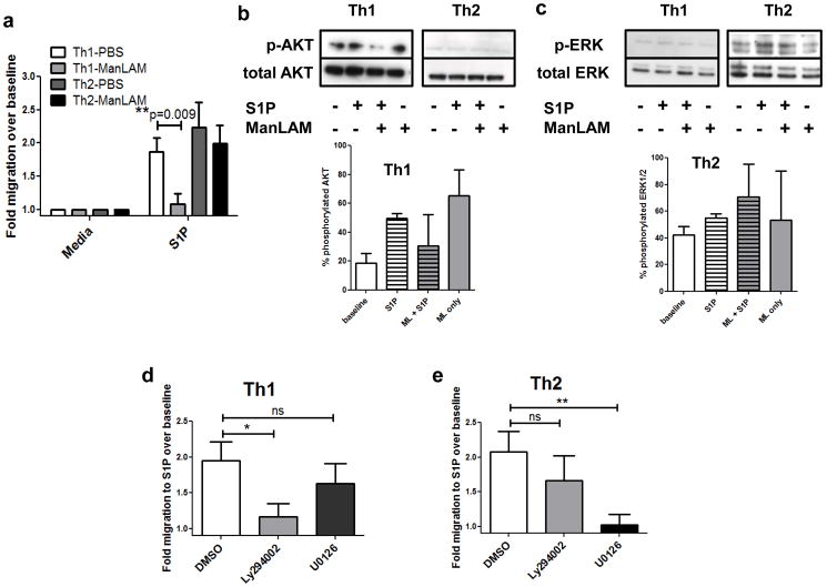
Figure 6. ManLAM inhibits Th1, but not Th2, cell migration induced by S1P because of differential signaling requirements.
(A) Spleen and lymph node T cells were isolated from 7–12wk old C57BL/6J female mice and skewed in vitro before treatment and Transwell assay. There was a statistically significant decrease in Th1 cell migration to 10nM S1P following 100ng/mL ManLAM treatment for 2h, and no significant difference in Th2 cell migration (n=3 experiments performed in triplicate, mean ± s.e.m., **P=0.009 Student’s t test). (B & C) Western blotting analysis of Th1 and Th2 cells following ManLAM pretreatment and S1P stimulation for (B) phosphorylated AKT or (C) phosphorylated ERK. Insets depict % phosphorylated protein averaged from blots from 2 separate experiments. (D) Th1 and (E) Th2 cell migration to 10nM S1P following a 1h treatment with DMSO vehicle, 10μM PI3K inhibitor Ly294002, or 10μM MEK1/2 inhibitor U0126. S1P-directed Th1 cell migration was inhibited by Ly294002 and S1P-directed Th2 cell migration was inhibited by U0126 pretreatment (n=3 separate experiments performed in triplicate, mean ± s.e.m., *P=0.02 and **P=0.005 Student’s t test).

