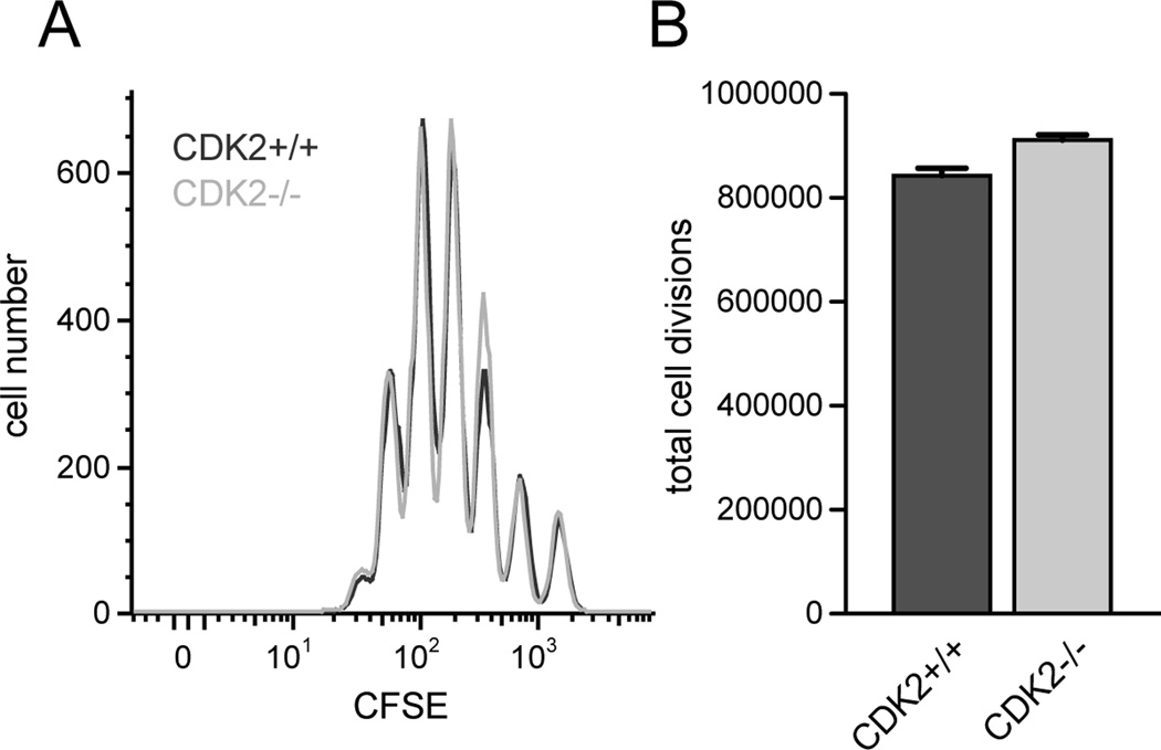
Figure 1.
Proliferation of CD4+ T cells from wild-type vs. CDK2-deficient mice. Purified CD4+CD25− T cells from wild-type (dark gray) or CDK2-deficient (light gray) mice were labeled with CFSE and stimulated in vitro with soluble anti-CD3 (0.5 µg/mL) in the presence of Thy1.2-depleted spleen cells for 72 hours, and cell division was assessed by flow cytometry (A). The total number of cell divisions achieved by each population was calculated as described previously (52) (B). The data depicted are representative of 4 independent experiments.

