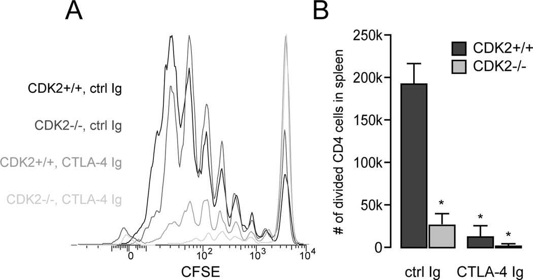
Figure 3.
In vivo mixed lymphocyte responses by wild-type vs. CDK2-deficient CD4+ T cells. Spleen and lymph node cells from wild-type or CDK2-deficient mice (H-2b) were labeled with CFSE, adoptively transferred into B6×DBA F1 recipients (H-2bxd), and the recipients were treated with CTLA-4Ig or control Ig. After 3 days, spleens were harvested and cell division by H-2d-negative donor CD4+ T cells was assessed by flow cytometry (A). The average number of divided CD4+ T cells (+/− SEM, p < 0.01) from three recipients per group is depicted in B and is representative of two independent experiments.

