Figure 5.
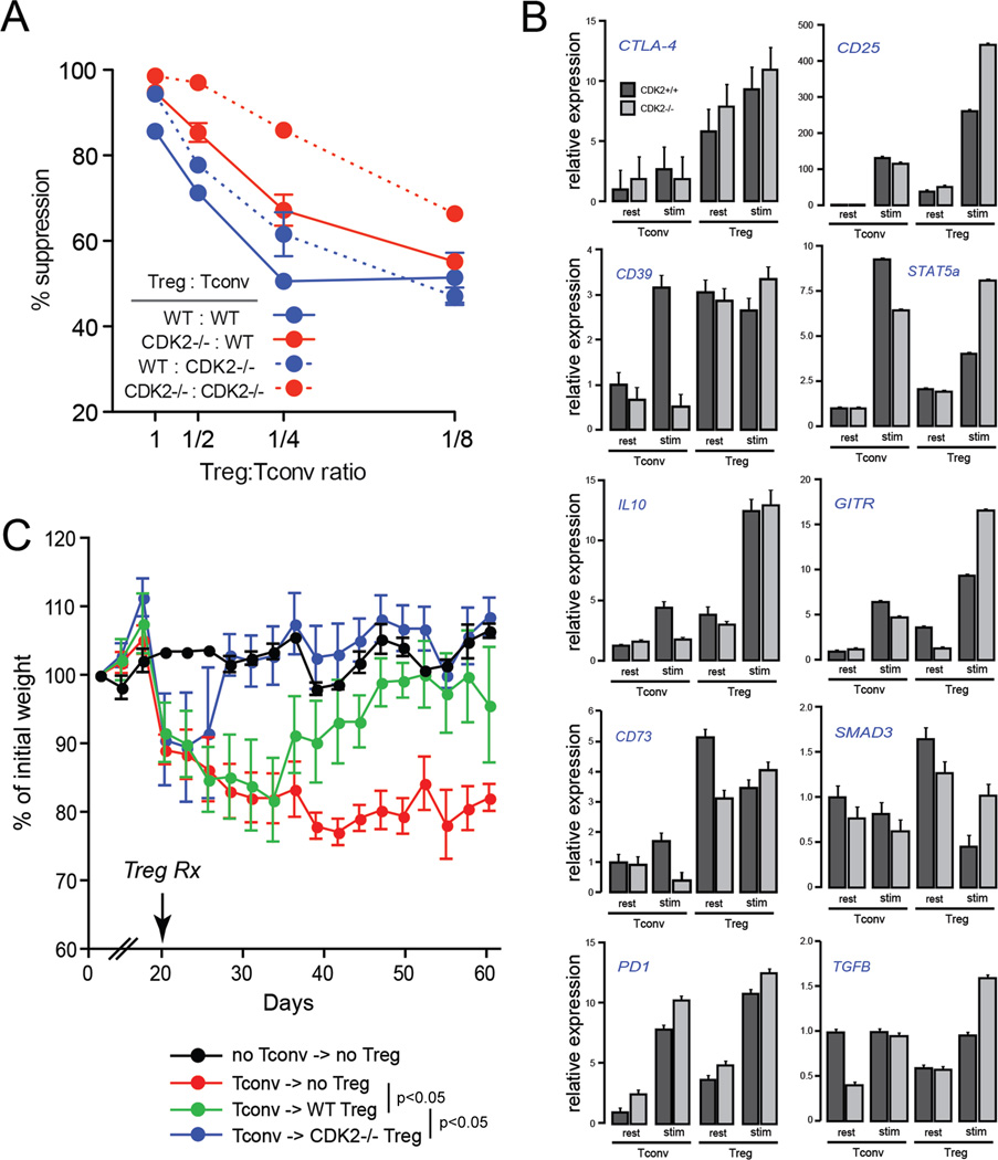
Regulatory T cell function in wild-type vs. CDK2-deficient mice. CD4+CD25+ Treg purified from naive wild-type or CDK2-deficient mice were cultured at different ratios with CD4+CD25− wild-type or CDK2-deficient Tconv in a standard anti-CD3-based in vitro suppression system. Plotted is the percent suppression of Tconv cell division as measured by CFSE (A). Gene expression by resting or 4-hour stimulated wild-type and CDK2-deficient Treg and Tconv was assessed by qRT-PCR (B). Naive CD4+CD25− T cells were purified from wild-type mice and adoptively transferred into Rag1-deficient recipients (C). At day 20 post-transfer, Rag1-deficient recipients (5 per group) received purified Treg from wild-type mice (green symbols), Treg from CDK2-deficient mice (blue symbols), or PBS as a control (red symbols). Recipients were subsequently weighed and monitored for symptoms of colitis every two days. The difference in weight kinetics between each treatment group was statistically significant (p < 0.05, ANOVA).

