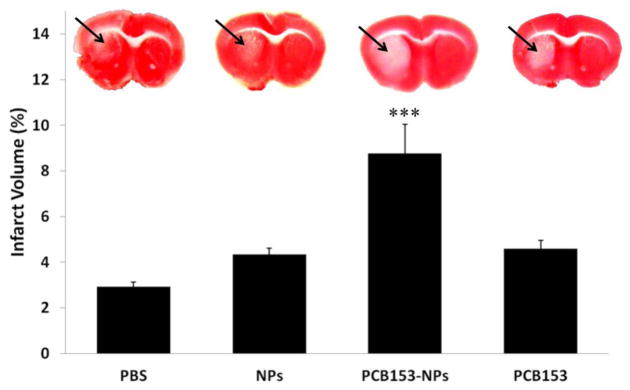
Figure 5. Exposure to PCB153-NPs increases the infarct volume in the experimental stroke model.
Mice were treated as in Figure 1 and subjected to a 40 min MCAO, followed by a 24 h reperfusion, and staining with 2,3,5-triphenyltetrazolium chloride (TTC) to visualize viable tissue. Unstained area (arrows) corresponds to damaged brain tissue. Quantified results of negative TTC staining are depicted in the form of bar graphs. Results are mean ± SEM, n=5–7. ***Significantly different as compared to other groups at p<0.001.

