
Figure 1.


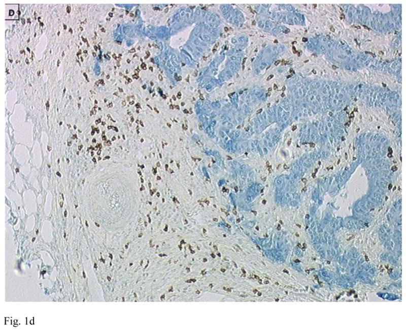
Immunohistology of primary colon tumors in a tissue microarray (TMA) showing examples of tumors with low (A and C) and high (B and D) CD8 T cell infiltration. Tumor is stained blue (cytokeratin) and CD8+ T cells brown. Images are courtesy of Dr. Jerome Galon.